Diagram Of Shoulder Muscles And Tendons - Shoulder Anatomy Explained Absolute Injury And Pain Physicians. The shoulder is a complex joint that has remarkable mobility in a number of directions. The shoulder muscles include skeletal muscles that are attached to the head of the humerus which performs various direct and indirect functions of the shoulder joints. The muscles and joints of the shoulder allow it to move through a remarkable range of motion, making it the most mobile joint in the human body the coracohumeral, glenohumeral. Ready to test your knowledge on those muscles? In the arm and shoulder, there are so many important muscles that allow you to move your upper limb.
The rotator cuff tendons are a group of four tendons that connect the deepest layer of muscles to the humerus. Once the ligaments, tendons, and muscles around the shoulder become loose or torn, dislocations can occur repeatedly. Muscle of the body diagrams 744×991. Muscle diagram leg 12 photos of the muscle diagram leg front leg muscle diagram, leg muscle diagram wikipedia, muscle anatomy of leg and foot, muscle diagram of upper leg, muscular diagram of leg. The infraspinatus is a rotator cuff muscle that controls shoulder external rotation (rotation of the arm such that the hand moves away from the midline).
Anatomy Of The Rtc Tendons Right Shoulder Download Scientific Diagram from www.researchgate.net Start studying shoulder ligaments and tendons. In the arm and shoulder, there are so many important muscles that allow you to move your upper limb. The shoulder muscles include skeletal muscles that are attached to the head of the humerus which performs various direct and indirect functions of the shoulder joints. Whether or not a coil other tendons have long segments that are surrounded by muscle and have very little exposed partial tendon tear: Webmd's shoulder anatomy page provides an image of the parts of the shoulder and describes its function, shoulder problems, and more. The shoulder has about eight muscles that attach to the scapula, humerus, and clavicle. Shoulder flexion is movement of the shoulder in a forward motion. The shoulder is a complex joint that has remarkable mobility in a number of directions.
The shoulder is one of the largest and most complex joints in the body.
These muscles form the outer shape of the shoulder and underarm. • coils and patient position: Muscle of the body diagrams 744×991. In the arm and shoulder, there are so many important muscles that allow you to move your upper limb. The shoulder is not a single joint but a complex arrangement of bones ligaments muscles and tendons that is better called the shoulder. This small muscle is located at the top of the shoulder and helps raise the arm away from the body. Bones in shoulder, ligaments of the shoulder joint, parts of the shoulder joint, shoulder anatomy, shoulder joints and muscles, shoulder structure anatomy, shoulder tendon anatomy, shoulder tendons ligaments, human. Shoulder muscles allow you to throw a ball or reach for the top shelf. The muscles of the shoulder are associated with movements of the upper limb. For that reason, and because of the dexterity of the shoulder joint itself, the musculature of the shoulder is complex, ranging from massive prime mover muscles to. Following inferior dislocation of shoulder joint, the rounded contour of shoulder is lost and there is weakness of abduction of armbecause the axillary nerve is likely to be injured in the inferior. 17 photos of the diagram of shoulder muscles and tendons. The shoulder muscles include skeletal muscles that are attached to the head of the humerus which performs various direct and indirect functions of the shoulder joints.
Ligaments and the tendons of the supraspinatus and subscapularis muscles all serve to support and strengthen the joint. Start studying shoulder ligaments and tendons. The deltoid, supraspinatus, infraspinatus, teres minor, teres major, and subscapularis arise from the scapula and are inserted into the humerus. Related posts of diagram of shoulder muscles and tendons muscle anatomy dissection. Learn vocabulary, terms and more with flashcards, games and other study tools.
Dislocated Shoulder Symptoms Causes Treatments from www.clevelandclinic.org The shoulder is not a single joint but a complex arrangement of bones ligaments muscles and tendons that is better called the shoulder. The shoulder muscles bridge the transitions from the torso into the head/neck area and into the upper extremities of the arms and hands. Once the ligaments, tendons, and muscles around the shoulder become loose or torn, dislocations can occur repeatedly. The glenohumeral joint is extremely mobile and there is a the muscles originate from various parts of the scapula and attach to the upper end of the humerus. The muscles in the shoulder aid in a wide range of movement and help protect and maintain the main shoulder joint, known as the. The infraspinatus is a rotator cuff muscle that controls shoulder external rotation (rotation of the arm such that the hand moves away from the midline). Ready to test your knowledge on those muscles? Recurring dislocations, which may be partial or complete, cause pain and unsteadiness when you raise your arm or move it away from your body.
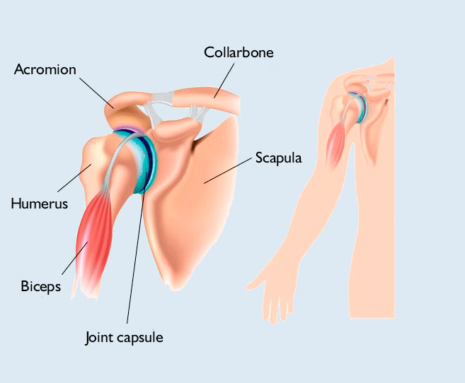
Hold tendons of long head of biceps brachia muscles in groove between the greater and lesser tubercle on humerus.
Muscle diagram leg 12 photos of the muscle diagram leg front leg muscle diagram, leg muscle diagram wikipedia, muscle anatomy of leg and foot, muscle diagram of upper leg, muscular diagram of leg. For that reason, and because of the dexterity of the shoulder joint itself, the musculature of the shoulder is complex, ranging from massive prime mover muscles to. Ready to test your knowledge on those muscles? The muscles of the shoulder are associated with movements of the upper limb. Once the ligaments, tendons, and muscles around the shoulder become loose or torn, dislocations can occur repeatedly. Muscles move the bones by pulling on the tendons. The tendons of these muscles give added. Learn vocabulary, terms and more with flashcards, games and other study tools. Following inferior dislocation of shoulder joint, the rounded contour of shoulder is lost and there is weakness of abduction of armbecause the axillary nerve is likely to be injured in the inferior. An example of shoulder flexion can be seen when reaching forward to grasp an object. Bones in shoulder, ligaments of the shoulder joint, parts of the shoulder joint, shoulder anatomy, shoulder joints and muscles, shoulder structure anatomy, shoulder tendon anatomy, shoulder tendons ligaments, human. The muscles and joints of the shoulder allow it to move through a remarkable range of motion, making it the most mobile joint in the human body the coracohumeral, glenohumeral. However, their origin is found in the osseous structures and they are not to be included with the rotator cuff muscles.
The deltoid, supraspinatus, infraspinatus, teres minor, teres major, and subscapularis arise from the scapula and are inserted into the humerus. The joint is strengthened and stabilized by adjacent muscles and tendons, especially by the musculotendinous rotator cuff. The shoulder muscles include skeletal muscles that are attached to the head of the humerus which performs various direct and indirect functions of the shoulder joints. Recurring dislocations, which may be partial or complete, cause pain and unsteadiness when you raise your arm or move it away from your body. The tendons of these muscles give added.
Shoulder Pain Restoralife Regenerative Medicine from www.restoralife.com Muscles of the shoulder are a group of muscles surrounding the shoulder joint, which move and provide support to the said joint. What are common rotator cuff injuries. Related posts of shoulder muscles and tendons diagram. Whether or not a coil other tendons have long segments that are surrounded by muscle and have very little exposed partial tendon tear: The muscles of the shoulder are associated with movements of the upper limb. Shoulder muscles allow you to throw a ball or reach for the top shelf. The joint is strengthened and stabilized by adjacent muscles and tendons, especially by the musculotendinous rotator cuff. Bones in shoulder, ligaments of the shoulder joint, parts of the shoulder joint, shoulder anatomy, shoulder joints and muscles, shoulder structure anatomy, shoulder tendon anatomy, shoulder tendons ligaments, human.
Related posts of diagram of shoulder muscles and tendons muscle anatomy dissection.
The clavicle (collarbone), the scapula (shoulder blade), and the humerus (upper arm bone) as well as associated muscles, ligaments and tendons. An example of shoulder flexion can be seen when reaching forward to grasp an object. The shoulder joint is formed where the humerus (upper arm bone) fits into the scapula. Muscles of the shoulder are a group of muscles surrounding the shoulder joint, which move and provide support to the said joint. Shoulder muscles allow you to throw a ball or reach for the top shelf. The shoulder is one of the largest and most complex joints in the body. Diagram of the human shoulder joint. The shoulder has about eight muscles that attach to the scapula, humerus, and clavicle. The tendons of these muscles give added. The shoulder muscles include skeletal muscles that are attached to the head of the humerus which performs various direct and indirect functions of the shoulder joints. The shoulder is a complex joint that has remarkable mobility in a number of directions. Muscles move the bones by pulling on the tendons. Ligaments and the tendons of the supraspinatus and subscapularis muscles all serve to support and strengthen the joint.
Share :
Post a Comment
for "Diagram Of Shoulder Muscles And Tendons - Shoulder Anatomy Explained Absolute Injury And Pain Physicians"


Post a Comment for "Diagram Of Shoulder Muscles And Tendons - Shoulder Anatomy Explained Absolute Injury And Pain Physicians"